
If your doctor has recommended a PSMA PET scan, you’re likely wondering what this test involves and why it’s different from other imaging studies. Let’s get into this advanced diagnostic tool in clear, practical terms.
What Is PSMA PET Scan?
A PSMA PET scan combines two technologies to create precise images of prostate cancer cells throughout your body. PSMA stands for Prostate-Specific Membrane Antigen, which acts like a unique address found primarily on prostate cancer cells. PET (Positron Emission Tomography) uses a small amount of radioactive material to highlight areas where cancer cells are most active.
Think of it this way: while a regular X-ray shows the structure of your bones, a PSMA PET scan specifically seeks out and illuminates prostate cancer cells, no matter where they might be hiding in your body.
How does the Scan Work?
The process begins with an injection of a radioactive tracer designed to attach specifically to PSMA proteins. This tracer acts like a guided missile that seeks out prostate cancer cells and sticks to them. Normal prostate tissue has some PSMA, but cancer cells typically have much higher concentrations, making them stand out clearly.
After receiving the injection through an IV, you’ll wait 60-90 minutes while the tracer circulates and accumulates where cancer cells are present. During the actual scan, you’ll lie on a table that moves through a large, donut-shaped scanner. The machine detects the radioactivity and creates detailed images showing exactly where the tracer has collected.
Types of PSMA Tracers
Different medical centers use various radioactive tracers, each with unique advantages. Gallium-68 PSMA provides excellent image quality and has been extensively studied, though it requires on-site production due to its short half-life. Fluorine-18 tracers offer more flexible scheduling and potentially better image quality in certain areas of the body.
Your doctor will select the most appropriate tracer based on your specific situation and what information they need to guide your treatment decisions.
Who Benefits from PSMA PET Scans?
These specialized scans are typically recommended for men with intermediate to high-risk prostate cancer to determine if the disease has spread beyond the prostate. This staging information dramatically influences treatment planning, helping doctors decide whether surgery, radiation, hormone therapy, or combination approaches would be most effective.
PSMA PET scans are also invaluable for patients whose PSA levels are rising after initial treatment. When conventional imaging fails to locate recurrent cancer, PSMA PET scans excel at finding hidden cancer cells, enabling targeted treatments rather than whole-body approaches.
Comparison of Different PET Scan Types
| Scan Type | Tracer Used | Best For | Advantages | Limitations |
| PSMA PET | PSMA-targeting tracers (68Ga-PSMA, 18F-DCFPyL) | Prostate cancer detection | Highly specific for prostate cancer. Detects small lesions (2-4mm). Changes treatment in 20-30% of cases | Not all prostate cancers express PSMA. More expensive. Requires specialized tracers |
| FDG PET | 18F-fluorodeoxyglucose (sugar-based) | Most other cancers | Widely available. Well-established. Good for many cancer types | Poor for prostate cancer. Less specific. High background activity |
| Conventional CT | Iodine contrast (if used) | Anatomy and large lesions | Excellent anatomical detail. Widely available. Cost-effective | Cannot specifically identify cancer. Misses small lesions. Limited functional information |
| Bone Scan | Technetium-99m | Bone metastases | Good for bone changes. Widely available. Cost-effective | Cannot distinguish cancer from other bone conditions. Less sensitive than PSMA PET. Limited to bone only |
| Multiparametric MRI | Gadolinium contrast | Prostate gland detail | Excellent soft tissue detail. No radiation. Good for local staging | Limited for detecting distant spread. Cannot specifically target cancer cells (Time-intensive) |
What to Expect During Your Scan?
Preparing for a PSMA PET scan is straightforward. Unlike some imaging tests, you can eat normally and continue taking your medications. Wear comfortable clothing without metal fasteners, and plan for a total appointment time of 2-3 hours.
On scan day, you’ll receive the radioactive tracer injection, which feels like any other IV insertion. During the waiting period, you can read, listen to music, or rest. The actual scanning takes 15-30 minutes while you lie still on a comfortable table.
The radioactive tracer contains only a tiny amount of radiation, similar to a CT scan, and most of it leaves your body within 24-48 hours through normal urination and radioactive decay.
Understanding Your Results
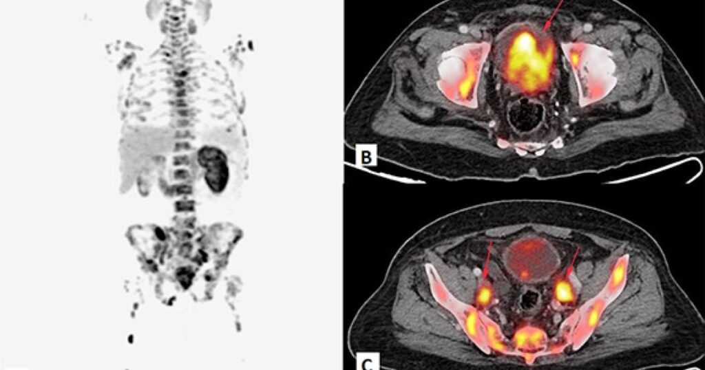
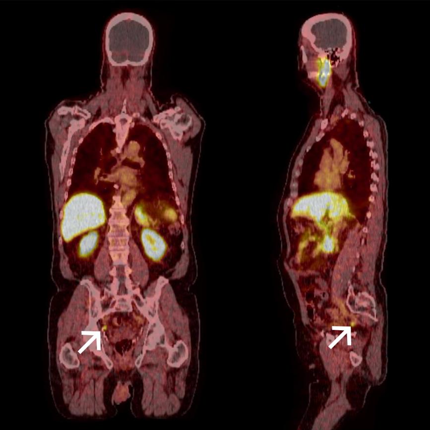
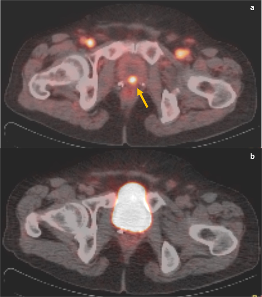
PSMA PET scan images show areas of tracer uptake as bright spots. Higher uptake typically indicates greater cancer activity, though experienced radiologists must distinguish between cancer and occasionally increased uptake from inflammation or other benign conditions.
Your scan report will detail the location, intensity, and number of any suspicious areas. This information helps determine whether cancer is confined to the prostate, has spread to nearby lymph nodes, or has reached distant sites like bones or organs.
Advantages Over Conventional Imaging
PSMA PET scans demonstrate superior sensitivity compared to traditional methods, detecting cancer deposits that CT scans or bone scans might miss. Studies show these scans change treatment recommendations in 20-30% of patients compared to conventional imaging alone.
This precision often allows for more targeted treatments. For example, if PSMA PET identifies specific areas of recurrence, doctors can focus radiation therapy on those exact locations rather than treating larger areas or using systemic therapy for the entire body.
Limitations to Consider
While highly accurate, PSMA PET scans aren’t perfect. Approximately 5-10% of prostate cancers have low PSMA expression, making them harder to detect. Very small cancer deposits might not accumulate enough tracer to be visible, and some aggressive cancers may lose PSMA expression over time, particularly after multiple treatments.
False positive results can occasionally occur when non-cancerous conditions cause increased PSMA uptake, though experienced radiologists can usually distinguish these from true cancer activity.
Impact on Treatment Decisions
PSMA PET results significantly influence treatment planning. For newly diagnosed patients, the scans help determine surgical candidacy and radiation therapy fields. When PSA levels rise after treatment, PSMA PET can identify recurrence locations, enabling targeted therapies rather than systemic treatments.
The precision of these scans sometimes allows treatment de-escalation when they show less extensive disease than conventional imaging suggested, while other times they reveal more widespread cancer requiring different therapeutic approaches.
Cost and Insurance Coverage
PSMA PET scans typically cost more than conventional imaging due to specialized equipment and tracers. However, insurance coverage has improved dramatically, with most major providers now covering these scans for appropriate clinical indications like staging intermediate to high-risk cancer or evaluating rising PSA after treatment.
Prior authorization may be required, and your healthcare team can help navigate the approval process by providing necessary clinical documentation.
Living with Your Results
Whether your scan shows clear results or areas of concern, remember that having accurate information enables your medical team to develop the most appropriate treatment strategy. Clear scans provide reassurance and support treatment planning, while positive results, though challenging emotionally, allow for targeted interventions using modern therapies that help many men maintain a good quality of life.
Consider connecting with support groups or counseling services if you’re struggling with your diagnosis or scan results. Many cancer centers offer these resources, and patient advocacy organizations provide valuable support networks.
Common Questions Answered
How does PSMA PET differ from regular PET scans?
Regular PET scans use sugar-based tracers that highlight general metabolic activity, while PSMA PET uses tracers specifically designed to attach to prostate cancer cells, making them much more sensitive and specific for prostate cancer detection.
Will I be radioactive after the scan?
You’ll have minimal radioactivity that decreases rapidly. Most facilities recommend avoiding close contact with pregnant women and young children for 6-24 hours, and the radioactivity returns to normal background levels by the next day.
Can the scan miss cancer?
While highly sensitive, PSMA PET scans can miss very small deposits or cancers with low PSMA expression. However, they still detect significantly more cancer than conventional imaging methods.
Why don’t all men with prostate cancer get these scans?
PSMA PET scans are reserved for situations where the information would change treatment decisions. Men with low-risk cancer on active surveillance typically don’t need this level of imaging.
Moving Forward
PSMA PET scans represent a significant advancement in prostate cancer care, providing unprecedented accuracy in detecting and staging the disease. Understanding this technology empowers you to participate actively in treatment decisions and reduces anxiety about the procedure itself.
Remember that your healthcare team uses these scans to provide the most effective treatment while maintaining your quality of life. Don’t hesitate to discuss any questions or concerns about PSMA PET scans with your medical team, as they can provide personalized guidance based on your specific situation.